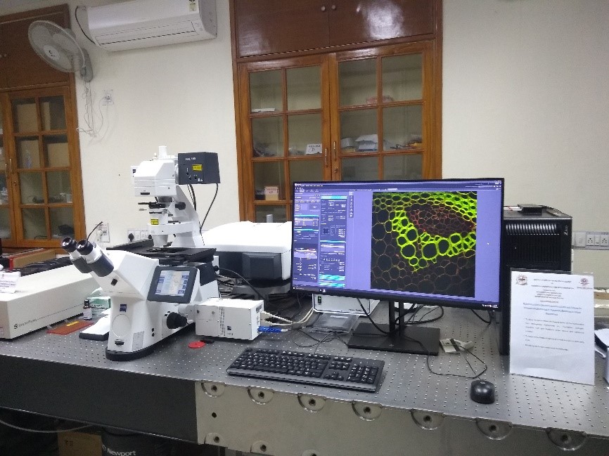
CONFOCAL MICROSCOPY LAB
- Department of Medical Physics has the facility of Confocal microscopy which is a powerful imaging technique that allows researchers and clinicians to view biological samples with high resolution and three-dimensional detail. The confocal microscopy can be used in various fields, including:
- 1. Biology and Life Sciences:
In biology and life sciences to study cell structure and function, cellular dynamics, and tissue architecture. It is particularly useful for studying fluorescently-labeled cells and molecules, such as tracking the movement of organelles within cells or studying the behavior of cancer cells.
- 2. Neuroscience:
In neuroscience to study the structure and function of the brain and nervous system. Researchers can use it to visualize and analyze the complex networks of neurons and synapses that underlie brain function, as well as to study the mechanisms of neurodegenerative diseases.
- 3. Material Science:
To study the structure and properties of materials, such as polymers, metals, and semiconductors. It can provide detailed images of the microstructure of materials, allowing researchers to analyze defects and other features that affect material properties.
- 4. Medical Applications:
In medical applications, including imaging of tissues and organs, as well as studying skin and hair. In dermatology, confocal microscopy is used to diagnose skin diseases and evaluate their severity. In ophthalmology, it can be used to image the retina and diagnose eye diseases.


